
Molecular workflow
Veterinary molecular testing: the mFIND workflow
See how mFIND moves from a prepared sample to a fluorescent signal, and what its 15-minute amplification stage means for workflow planning.
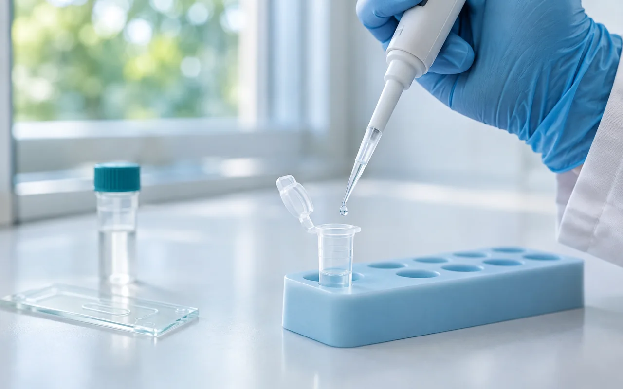
Veterinary molecular testing detects defined nucleic acid targets. In Dawnsail’s current mFIND explanation, the workflow combines sample preparation, distribution into a sealed microfluidic chip and constant-temperature amplification with fluorescence monitoring. The documented 15-minute figure refers to the amplification stage, rather than the complete time from collecting a specimen to reviewing a report.
For a laboratory evaluating mFIND, the useful starting point is to map the full sequence. This shows which steps involve an operator, which take place inside the chip and which details must be confirmed for the selected assay.
The workflow below follows the current veterinary mFIND materials and Dawnsail’s approved principle explanation. It is a process overview, rather than a replacement for the assay instructions or an operating protocol.
What does isothermal amplification mean here?
Isothermal amplification uses a constant reaction temperature. Dawnsail’s supplied mFIND principle describes four enzymes working together at 40°C, with the analyzer monitoring fluorescence as amplification proceeds. The current explanation does not identify the enzymes individually; this guide therefore stays with the documented method description. 1
This is a more precise description of mFIND than calling it a conventional thermal-cycling PCR instrument. Both approaches belong to molecular testing, but an assay name, reaction protocol or interpretation rule from one system cannot simply be transferred to the other.
If you are comparing molecular detection with biomarker measurement, our immunoassay versus molecular testing guide explains the difference in targets and recognition methods.
How does the sample move through mFIND?
1. Prepare the sample using the specified buffers
The supplied workflow describes adding the sample to buffer A to release nucleic acid, then transferring the sample solution to buffer B to stabilize it. These are the functions stated in the current product explanation. Exact specimen requirements, volumes and handling conditions belong to the selected assay’s instructions. 1
When planning a demonstration, identify those instructions first. A respiratory panel and a digestive panel should not be assumed to accept the same specimen or preparation procedure simply because both use mFIND.
2. Load, seal and distribute the sample
The prepared solution is added to the chip’s sample well and the chip is sealed. The current explanation then describes centrifugation distributing the solution through the channel into four reaction chambers. Amplification takes place within the sealed chip. 1
The number of chambers describes the supplied chip structure. It does not, by itself, tell a buyer how many reportable pathogens a particular panel includes. Confirm the target list and any control allocation in the documentation for that panel.
For an implementation review, ask the supplier to show the required loading and centrifugation equipment, the approved consumables and the complete handling sequence. Record which items are supplied with the selected configuration.
3. Amplify and monitor fluorescence
The documented amplification stage runs at 40°C for 15 minutes. Dawnsail’s principle explanation states that the analyzer excites and reads fluorescence every 10 seconds, providing a view of the amplification process, and displays a positive signal when the applicable threshold is reached. 1
These figures describe the supplied process. They are not a universal performance claim for every molecular assay, and a fluorescence threshold is not automatically a calibrated pathogen concentration. Review the actual report format and interpretation rules for the selected panel.
Swipe horizontally to read the full table.
| Workflow stage | What the current mFIND explanation describes | Information to confirm before implementation |
|---|---|---|
| Sample preparation | Buffer A releases nucleic acid; buffer B stabilizes it | Accepted specimen, preparation requirements and materials |
| Chip loading | Sample solution enters the sample well; the chip is sealed | Loading procedure, consumables and sealing checks |
| Distribution | Centrifugation distributes solution into four chambers | Required equipment, panel targets and control allocation |
| Amplification | Constant-temperature reaction at 40°C for 15 minutes | Applicable assay program and documented run requirements |
| Signal and report review | Fluorescence is monitored during amplification | Result categories, validity checks and follow-up instructions |
How should you interpret the 15-minute figure?
Use amplification time and total workflow time as separate fields in an evaluation sheet. The first describes a defined instrument stage. The second includes everything needed to reach a reviewed result in the laboratory’s actual process.
A useful workflow map includes specimen collection or receipt, preparation, loading and sealing, distribution, amplification, and result review. If a specimen has to wait for an operator or equipment, that waiting time also affects the laboratory’s turnaround.
During a demonstration, record the start and finish of each stage. This produces a useful local observation without turning a single timed run into a claim about every specimen or operating condition. Ask the team to distinguish hands-on time from time during which the instrument runs independently.
What does a sealed chip change?
The sealed-chip format keeps the described amplification reaction inside a small enclosed space. Dawnsail presents this as part of its approach to limiting aerosol contamination during the reaction. It does not establish that sample preparation, loading or the entire laboratory environment is free from contamination risk. 1
For planning purposes, ask how the documented workflow separates specimen handling, reagent preparation and used consumables. Review cleaning, waste handling and unexpected-result procedures with the supplier. Do not infer a specific room layout or biosafety requirement from the chip format alone; the laboratory’s assessment and the applicable instructions determine those requirements.
What should you review before accepting a result?
Start with the selected assay’s validity criteria and report definitions. Cornell’s molecular diagnostic laboratory describes using amplification and extraction controls, with quality criteria that must be satisfied before reporting its PCR results. This illustrates the role of controls in molecular testing; it does not describe the control configuration of an mFIND chip. 2
For mFIND, request the documented answers to these questions:
- Which controls apply to the selected panel, and how are they represented in the report?
- What distinguishes a valid negative result from an invalid run?
- What action is specified when a control, loading check or instrument check fails?
- Which result categories and flags can appear, and which written rules govern them?
Also match performance evidence to the intended use. For infectious-disease assays, WOAH’s guidance calls for validation in the relevant species and specimen. A result from one evaluated panel or specimen cannot establish the performance of every test offered on an instrument. 3
Which documents should a distributor request?
Prepare a package for the actual assays being discussed: the current instrument manual, each panel’s instructions, the target and specimen list, the applicable quality-control procedure and the relevant validation information. Add the documented equipment list and training materials so the laboratory can review the complete workflow.
The Dawnsail resource library provides platform information and available manuals. The mFIND product page is the starting point for reviewing the veterinary test menu. Use these together to create a shortlist, then contact Dawnsail with the required targets, animal species and intended market.
Common questions
Do four reaction chambers mean four different pathogens?
Not necessarily. Chamber count describes chip architecture. The assay documentation must identify the reportable targets and any control allocation for the selected panel.
Does a positive fluorescence signal give a pathogen concentration?
The current principle text describes a threshold-based positive signal. It does not establish a calibrated quantitative result. Use the output and interpretation defined for the individual assay.
Can the whole workflow be advertised as 15 minutes?
The supplied text supports a 15-minute amplification stage. Collection, preparation, loading, distribution and review must be accounted for separately when describing the complete workflow.
Sources and product documents
Product details follow Dawnsail’s October 2026 approved mFIND principle explanation. The external references explain general control and validation principles; they are not independent performance evaluations of mFIND.
- Dawnsail — mFIND principle and supporting materials. Buffers, sealed-chip distribution, reaction temperature, amplification duration and fluorescence monitoring.
- Cornell University — Interpretation of PCR results. Assay-specific reporting and the role of quality controls in its PCR laboratory.
- WOAH — Validation of Diagnostic Assays for Infectious Diseases of Terrestrial Animals (PDF). Validation for an intended purpose, species and specimen.